All-optical photoacoustic imaging for neurobiology
Light provides rich molecular contrast, but it rapidly loses direction in brain tissue. Ultrasound travels much farther with little scattering. Photoacoustic imaging combines these properties: short laser pulses generate ultrasound inside tissue, and the recorded waves reveal where light was absorbed.
Our long-term goal is to image neuronal activity non-invasively beyond one millimetre depth, with resolution and sensitivity relevant to neural circuits. The project brings together transparent ultrasound sensors, fast optical interrogation and three-dimensional reconstruction, developed as parts of a single imaging system.
Why photoacoustics can reach deeper
A nanosecond laser pulse briefly heats absorbing structures such as blood vessels or functional indicators. Their thermoelastic expansion emits broadband pressure waves. Because these waves are only weakly scattered by soft tissue, they can be measured at the surface and used to recover a three-dimensional map of optical absorption.
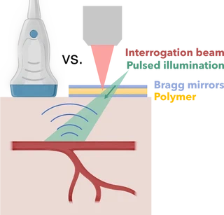
Photoacoustic imaging combines optical absorption contrast with acoustic propagation at depth. Temporary figure from the Institut Fresnel project page.
Our contribution: measuring sound with light
We develop transparent Fabry-Pérot ultrasound sensors made from a thin polymer layer between dielectric mirrors. An incoming pressure wave changes the layer thickness by a tiny amount and shifts its optical resonance. A focused interrogation beam converts this shift into a measurable change in reflected light.
The thin sensing layer provides broad acoustic bandwidth and high-frequency sensitivity, both of which are needed to resolve small structures. Scanning the interrogation beam across the sensor records the acoustic field over space and time; an inverse model then reconstructs the original absorber distribution.
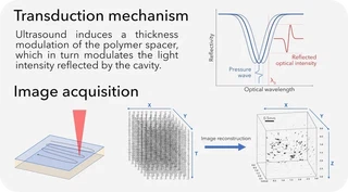
Optical ultrasound detection and raster-scanned acquisition. Temporary figure from the Institut Fresnel project page.
Current technical work
Small thickness variations across a fabricated sensor shift its resonance from point to point. A conventional narrowband system must therefore retune its wavelength during a scan, which limits acquisition speed. We are developing several ways around this limitation: broadband illumination with fast wavelength selection, dual-comb optical interrogation that samples many wavelengths at once, and acquisition strategies that require fewer spatial measurements.
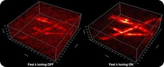
Compensating resonance inhomogeneity recovers image quality across the sensor. Temporary figure from the Institut Fresnel project page.
Fast three-dimensional reconstruction
Accurate model-based reconstruction can include detector physics, acquisition geometry, noise and prior knowledge, but repeatedly solving the full acoustic wave equation is expensive. We are developing fast forward and adjoint operators tailored to planar detection so that iterative three-dimensional reconstruction becomes practical.
From instrumentation to neurobiology
The instrumentation is being developed toward functional imaging of neuronal and vascular signals. This requires connecting sensor performance, illumination, reconstruction and biological validation rather than optimizing any one component in isolation. We work with neurobiology collaborators at INMED and INT on relevant questions and preparations.
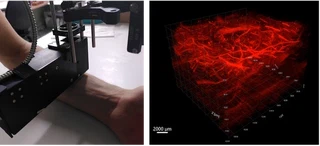
An experimental system and an example vascular reconstruction. Temporary figure from the Institut Fresnel project page.
Working with us
This programme sits at the interface of optical instrumentation, thin-film sensing, inverse problems and neurobiology. Collaborations can contribute a strong biological question, a new functional contrast mechanism or complementary expertise in imaging and reconstruction. Students and postdoctoral researchers may work across experiment, sensor development and quantitative image formation; contact us to discuss a possible fit.
Selected sources
Institut Fresnel: All-optical photoacoustic imaging. J. Saucourt et al., Fast interrogation wavelength tuning for all-optical photoacoustic imaging, Optics Express 31, 11164–11177 (2023). E. Küçükkomürcü et al., A depth-dependent, transverse shift-invariant operator for fast iterative 3D photoacoustic tomography in planar geometry (2026 preprint).
