Photoacoustic-guided wavefront shaping
Coherent light becomes a granular speckle pattern after propagating through a strongly scattering material. Although the original beam appears to have lost its direction, the propagation remains deterministic over a short time. By controlling the phase of the incident light, it is therefore possible to make the scattered waves interfere constructively at a chosen position.
This principle is well established when the target can be observed with a camera. Focusing inside biological tissue is harder because the target is hidden and an internal source of feedback is required. We investigate whether a photoacoustic signal—generated only where light is absorbed—can provide that guide from within the tissue.
Photoacoustics as an internal guide star
Photoacoustics provides feedback from absorbing structures below the surface. A spatial light modulator changes the incident optical wavefront, the absorber converts the local optical energy into an ultrasonic signal, and an ultrasound detector measures the result. The phase pattern can then be optimized to increase the signal from a selected acoustic region.
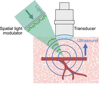
A spatial light modulator controls the incident field, while the photoacoustic signal provides feedback from inside the scattering medium. Adapted from Xia et al. (2024).
An alternative is to measure a photoacoustic transmission matrix. In this approach, the elements of a photoacoustic image take the place of camera pixels and record how a set of input optical modes influences absorbing targets. Once measured, the matrix can be used to calculate the wavefront that concentrates light on a selected target.
What has been demonstrated
Early experiments demonstrated this approach through scattering samples, including 0.5 mm of chicken tissue, and extended it to two-dimensional photoacoustic imaging with a linear ultrasound array. These results established non-invasive acoustic feedback as a practical route to optical control behind a scattering layer.
Why deep tissue remains difficult
The remaining difficulty is the large difference between optical and acoustic length scales. A single acoustically resolved volume may contain thousands of independent optical speckles. Increasing the total photoacoustic signal from that volume does not automatically produce one sharply focused optical spot, and the useful modulation can be very small compared with the background signal.
Deep biological tissue also decorrelates as it moves, so the wavefront must be measured and updated before the scattering pattern changes. Detector sensitivity, laser stability, dynamic range and the speed and number of modes of the spatial light modulator all become limiting factors. As reviewed by Xia et al. (2024), concentrating light deep inside living tissue has therefore not yet been demonstrated in the same way as focusing through static scattering layers.
Current direction
We study how photoacoustic feedback can be made more selective and more rapidly acquired, and how optical modulation, acoustic detection and reconstruction should be designed together. The aim is both to approach optical focusing at depth and to develop less stringent forms of light redistribution that can improve photoacoustic excitation or fluorescence measurements in scattering tissue.
Working with us
Progress depends on combining wavefront control, high-sensitivity ultrasound detection, fast optimization and realistic biological samples. The project is well suited to collaborators, students and postdoctoral researchers interested in the boundary between optical physics, acoustics and computational imaging. Contact us if you would like to explore a shared experiment or methodological question.
References
F. Xia, I. Leite, R. Prevedel and T. Chaigne, Optical wavefront shaping in deep tissue using photoacoustic feedback, Journal of Physics: Photonics (2024). T. Chaigne, Control of scattered coherent light and photoacoustic imaging: toward light focusing in deep tissue and enhanced, sub-acoustic resolution photoacoustic imaging, doctoral thesis, Université Paris 6 (2016).
